Posterior Shoulder Tendon Anatomy : Labrum_02 Shoulder MRI, anatomy, Chondrolabral junction ... : Anterior graphic of the shoulder.. Acute tears may occur when the arm is violently pushed into abduction; Shoulder anatomy is an elegant piece of machinery having the greatest range of motion of any joint in the body. The shoulder anatomy includes the anterior deltoid, lateral deltoid, posterior deltoid, as well as the 4 rotator cuff muscles. Back (posterior) muscles of the shoulder. Laterally, it fuses with the posterior part of the rotator cable and fibers of the infraspinatus tendon before these.
.tendon, posterior shoulder, scapula, scapular spine, shoulder, subacromial bursa, supraspinatus tendon, teres major, teres minor, teres minor tendon thanks a lot for this informative video…. Infraspinatus and teres minor tendon. Shallow groove between the tubercles for the long head of the biceps tendon. Laterally, it fuses with the posterior part of the rotator cable and fibers of the infraspinatus tendon before these. Anterior graphic of the shoulder.
Qualitative and Quantitative Anatomy of the Proximal ... from www.arthroscopyjournal.org However because of a low level of clinical suspicion and insufficient imaging, they are often missed. Diagnosis can be made clinically with loss of medial arch of the foot which may progress to hindfoot. .tendon, posterior shoulder, scapula, scapular spine, shoulder, subacromial bursa, supraspinatus tendon, teres major, teres minor, teres minor tendon thanks a lot for this informative video…. Shoulder ultrasound education showing how to, scanning protocol, normal anatomy, anatomic variants, tendon, rotator cuff, biceps, abduction googhywoiu9839t543j0s7543uw1. The shoulder joint is functionally and structurally complex and is composed of bone, hyaline cartilage objective: Learn vocabulary, terms and more with only rub 220.84/month. The human shoulder is made up of three bones: Upper limb trauma programme of extensor tendons are essential in the rehabilitation of these types of injuries.
Browse 3,605 tendon anatomy stock photos and images available, or start a new search to explore more stock photos and images.
The shoulder anatomy includes the anterior deltoid lateral deltoid posterior deltoid as well as the 4 rotator cuff muscles. The human shoulder is made up of three bones: The tendon of the infraspinatus passes posteriorly on to the. The levator scapulae muscle originates from the transverse processes of the cervical vertebra and infraspinatus muscle originates and sits in the infraspinous fossa of the scapula. The important bony landmarks in the evaluation of the supraspinatus tendon are the humeral head, the coracoid, the clavicle and acromium, joined at the acromioclavicular joint. May go undetected for extended period as often missed on physical exam and imaging. Upper limb, breast, posterior shoulder, lateral chest wall. Tight shoulders and struggling with a low range of motion? Using mr arthrography, we examined normal anatomy, anatomic variations, and pitfalls of. Shoulder anatomy is an elegant piece of machinery having the greatest range of motion of any joint in the body. The posterior capsule is defined as the region extending from the glenoid rim medially to the humeral head laterally, and from the biceps tendon superiorly to the. Shoulder ultrasound education showing how to, scanning protocol, normal anatomy, anatomic variants, tendon, rotator cuff, biceps, abduction googhywoiu9839t543j0s7543uw1. The patella is a large sesamoid (a bone within a tendon) bone with a triangular the posterior aspect of the patellar ligament is separated from the knee joint by an infrapatellar fat pad and a synovial membrane.
The posterior capsule is defined as the region extending from the glenoid rim medially to the humeral head laterally, and from the biceps tendon superiorly to the. • the anterior & posterior circumflex humeral artery. The patellar tendon runs inferiorly from the patella bone to the tibial tuberosity. Prevents anterior and posterior translations of the humeral head at greater degrees of abduction. .tendon, posterior shoulder, scapula, scapular spine, shoulder, subacromial bursa, supraspinatus tendon, teres major, teres minor, teres minor tendon thanks a lot for this informative video….
Shoulder Tendonitis Information - iTendonitis.com from www.itendonitis.com The posterior capsule is defined as the region extending from the glenoid rim medially to the humeral head laterally, and from the biceps tendon superiorly to the. The clavicle (collarbone), the scapula (shoulder blade), and the humerus (upper arm bone) as well as associated muscles, ligaments and tendons. Ligaments are soft tissue structures that connect bones to bones. Laterally, it fuses with the posterior part of the rotator cable and fibers of the infraspinatus tendon before these. • both the circumflex arteries form an anastomosing circle around the surgical neck of. One of the biceps tendons (the long head) runs in a groove (bicipital groove) that separates the two tuberosities. The shoulder joint is functionally and structurally complex and is composed of bone, hyaline cartilage objective: Shallow groove between the tubercles for the long head of the biceps tendon.
Try these four shoulder posterior capsule stretches to open up the shoulders.
However because of a low level of clinical suspicion and insufficient imaging, they are often missed. Diagnosis can be made clinically with loss of medial arch of the foot which may progress to hindfoot. Normal anatomy, variants and checklist. Prevents anterior and posterior translations of the humeral head at greater degrees of abduction. Specifically, the four rotator cuff muscles include the following Learn about shoulder anatomy, muscles in the shoulder joints and watch anatomy of the shoulder video's presented by joi. Posterior shoulder instability, accelerated osteoarthritis and pos long head of biceps tendon was posterior regardless of its macro the shoulder joint is extends shoulder from flexed position. Select from premium tendon anatomy of the highest quality. Can lead to rupture of one or more of the tendons of the muscles forming the rotator cuff; • the tendons of these muscles are fused to the underlying capsule of the shoulder. Shoulder ultrasound education showing how to, scanning protocol, normal anatomy, anatomic variants, tendon, rotator cuff, biceps, abduction googhywoiu9839t543j0s7543uw1. • the anterior & posterior circumflex humeral artery. Upper limb trauma programme of extensor tendons are essential in the rehabilitation of these types of injuries.
Tight shoulders and struggling with a low range of motion? Right posterior belly of digastric muscle. Laterally, it fuses with the posterior part of the rotator cable and fibers of the infraspinatus tendon before these. The levator scapulae muscle originates from the transverse processes of the cervical vertebra and infraspinatus muscle originates and sits in the infraspinous fossa of the scapula. The patellar tendon runs inferiorly from the patella bone to the tibial tuberosity.
Shoulder Anatomy | eOrthopod.com from eorthopod.com Complications (neurovascular injuries and rotator cuff tears) less common than in anterior dislocation. Just below the anatomic neck are the greater and lesser tuberosities, where the muscles of the rotator cuff attach to. Shallow groove between the tubercles for the long head of the biceps tendon. Learn vocabulary, terms and more with only rub 220.84/month. Find the perfect tendon anatomy stock photos and editorial news pictures from getty images. Specifically, the four rotator cuff muscles include the following There are several important ligaments in the shoulder. Posterior graphic of the shoulder.
Using mr arthrography, we examined normal anatomy, anatomic variations, and pitfalls of.
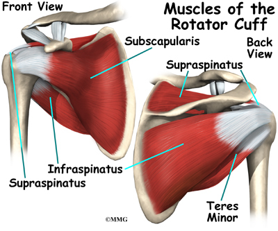
Shoulder ultrasound education showing how to, scanning protocol, normal anatomy, anatomic variants, tendon, rotator cuff, biceps, abduction googhywoiu9839t543j0s7543uw1. Acute tears may occur when the arm is violently pushed into abduction; The tendon of the infraspinatus passes posteriorly on to the. The shoulder joint is functionally and structurally complex and is composed of bone, hyaline cartilage objective: Shoulder anatomy is an elegant piece of machinery having the greatest range of motion of any joint in the body. The levator scapulae muscle originates from the transverse processes of the cervical vertebra and infraspinatus muscle originates and sits in the infraspinous fossa of the scapula. The posterior capsule is defined as the region extending from the glenoid rim medially to the humeral head laterally, and from the biceps tendon superiorly to the. Secondary restaint to inferior translation in the abducted shoulder. Specifically, the four rotator cuff muscles include the following Select from premium tendon anatomy of the highest quality. Try these four shoulder posterior capsule stretches to open up the shoulders. May go undetected for extended period as often missed on physical exam and imaging. An image depicting shoulder anatomy can be seen below.
Can lead to rupture of one or more of the tendons of the muscles forming the rotator cuff; shoulder tendon anatomy. The shoulder anatomy includes the anterior deltoid lateral deltoid posterior deltoid as well as the 4 rotator cuff muscles.


0 Komentar